Basic HTML Version


104
Escarpment Magaz ine Spr ing 2012
IN CONVERSATION
escarpment
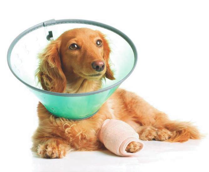
FOUR LEGGED FRIENDS
By DR. JACQUIE PANKATZ
Like
humans, dogs and cats have an anterior and a posterior cruciate ligament that criss-cross in the knee
providing stabilization of the joint. The anterior cruciate ligament is the one most commonly involved in
rupture in dogs. Whenever a pet, especially a large breed dog, presents with sudden hind leg limping, an-
terior cranial cruciate rupture is a possibility. These pets may have had a history of injury or playing or running
hard when all of a sudden there is a yelp and then a hind leg is lifted up. They may then become totally non-
weight bearing on the affected leg or walk with a dramatic limp and tend to only toe touch on the affected
side when standing. Sometimes they will sit in position such that the affected hind leg is stretched out as they
are hesitant to bend the leg. Others may present with a more chronic history of waxing and waning hind
limb limping that does not go away with rest or anti-inflammatory treatment. These pets may have a partial
tear of the ligament but often complete tear will develop over time.
Anterior cranial cruciate rupture is one of the most common canine orthopedic conditions seen in veterinary
practice. Any size of breed of dog can tear their cruciate ligament but large breed dogs appear to be more
commonly affected. One study showed that the Neapolitan mastiff, Newfoundland, Akita, St. Bernard, Rot-
tweiler, Chesapeake Bay retriever, and the American Staffordshire terrier are higher at risk for cruciate rup-
ture. In our veterinary practice, Labrador retrievers, Golden retrievers and their crosses also appear to be
commonly affected.
Older large dogs, especially if overweight, can have weakened ligaments and slowly stretch or partially
tear them. The partial rupture may be detected or the problem may not become apparent until the ligament
breaks completely. In this type of patient, stepping down off the bed or a small jump can be all it takes to
break the ligament.
A diagnosis of anterior cranial cruciate repair is often made after your veterinarian reviews the history, per-
forms an orthopedic exam and takes x-rays of the hind limbs. Because other disease conditions can also pro-
duce similar clinical signs, careful palpation of the knee as well as review of x-rays will greatly aid in making
the proper diagnosis. Many patients are best examined under sedation so that an abnormal movement,
known as a cranial drawer sign, can be detected. Other conditions that can mimic cruciate tears include
hip dysplasia and arthritis, displacement of the knee cap, fractures of the tip of the tibial bone and bone or
joint cancers. X-rays of the hips and both knee joints also screens patients for arthritis that may be present
secondary to the injured cruciate ligament.
Treatment of anterior cruciate tears requires surgical intervention. The goal of surgical repair is to stabilize
the knee so that the pet can regain normal function again and to slow the process of progressive degenerative
joint disease or arthritis that sets in when a rupture has occurred. It is important to realize, however, that there
are often already some degenerative joint changes that can be present by the time a diagnosis is made, and
the knee will likely develop arthritic changes as the pet ages. Affected patients are usually managed with
anti-inflammatories if needed and joint protective supplements along with weight-management and proper
diet. The recent development of veterinary formulated joint diets has been a valuable tool in helping manage
pets with osteoarthritis. Newer therapies including acupuncture, therapeutic laser and underwater treadmills
are also increasingly being utilized in veterinary medicine to manage these patients.
Canine Cranial
Cruciate Disease